
Antibody and Dye Based Assays

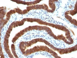
Biotium Cytokeratin 19(Ks19.1), CF488A conjugate, 0.1mg/mL
This MAb reacts with the rod domain of human cytokeratin-19 (CK19), a polypeptide of 40 kDa. Its epitope maps between amino acid 312-335. CK19 is expressed in sweat gland, mammary gland ductal and secretory cells, bile ducts, gastrointestinal tract, bladder urothelium, oral epithelia, esophagus, and ectocervical epithelium. Anti-CK19 reacts with a wide variety of epithelial malignancies including adenocarcinomas of the colon, stomach, pancreas, biliary tract, liver, and breast. Perhaps the most useful application is the identification of thyroid carcinoma of the papillary type, although 50%-60% of follicular carcinomas are also labeled. Anti-CK19 is a useful marker for detection of tumor cells in lymph nodes, peripheral blood, bone marrow and breast cancer.Primary antibodies are available purified, or with a selection of fluorescent CF Dyes and other labels. CF Dyes offer exceptional brightness and photostability. Note: Conjugates of blue fluorescent dyes like CF405S and


Biotium CD20(109-3C2), CF488A conjugate, 0.1mg/mL
Recognizes a protein of 30-33 kDa, which is identified as CD20 (Workshop V; Code CD20.12. Workshop IV; Code B17). It recognizes an extracellular domain of CD20. It is a non-Ig differentiation antigen of B-cells and its expression is restricted to normal and neoplastic B-cells, being absent from all other leukocytes and tissues. CD20 is expressed by pre B-cells and persists during all stages of B-cell maturation but is lost upon terminal differentiation into plasma cells. The protein passes through the membrane 4 times with both ends in cytoplasm and exposes one short and one longer loop to the external environment. CD20 is not glycosylated in resting B-cells and its cytoplasmic domains are differentially phosphorylated upon activation. It acts as calcium channel involved in B cell activation and cell cycle progression.Primary antibodies are available purified, or with a selection of fluorescent CF Dyes and other labels. CF Dyes offer exceptional brightness and photostabilit
Biotium CD7(B-F12), CF488A conjugate, 0.1mg/mL
Recognizes a protein of 40 kDa, identified as CD7 (also known as gp40, Leu9). CD7 is a member of the immunoglobulin gene superfamily. Its N-terminal amino acids 1-107 are highly homologous to Ig kappa-L chains whereas the carboxyl-terminal region of the extracellular domain is proline-rich and has been postulated to form a stalk from which the Ig domain projects. CD7 is expressed on the majority of immature and mature T-lymphocytes, and T cell leukemia. It is also found on natural killer cells, a small subpopulation of normal B cells and on malignant B cells. Cross-linking surface CD7 positively modulates T cell and NK cell activity as measured by calcium fluxes, expression of adhesion molecules, cytokine secretion and proliferation. CD7 associates directly with phosphoinositol 3'-kinase. CD7 ligation induces production of D-3 phosphoinositides and tyrosine phosphorylation.Primary antibodies are available purified, or with a selection of fluorescent CF Dyes and other labels.
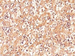
Biotium AFP (Alpha Fetoprotein)(C3), CF488A conjugate, 0.1mg/mL
Alpha-fetoprotein (AFP) is an oncofetal glycoprotein with a single chain of 70 kDa. This MAb is highly specific to AFP and shows no cross-reaction with other oncofetal antigens or serum albumin. AFP is normally synthesized in the liver, intestinal tract, and yolk sac of the fetus. Antibody to AFP has been shown to be useful in detecting hepatocellular carcinomas (HCC) and germ cell neoplasms, especially yolk sac tumors.Primary antibodies are available purified, or with a selection of fluorescent CF Dyes and other labels. CF Dyes offer exceptional brightness and photostability. Note: Conjugates of blue fluorescent dyes like CF405S and CF405M are not recommended for detecting low abundance targets, because blue dyes have lower fluorescence and can give higher non-specific background than other dye colors.
Biotium HER-2 / CD340(HRB2/258), CF488A conjugate, 0.1mg/mL
Recognizes a protein of 185 kDa, which is identified as c-erbB-2/HER-2/neu. Its epitope is localized in the extracellular domain. C-erbB-2/HER-2 is a member of the EGFR family. This MAb is specific and shows minimal cross-reaction with other members of the EGFR-family. Receptors of this family are located on the plasma membrane and consist of an extracellular ligand-binding domain that is connected to a large intracellular domain by a single transmembrane sequence. C-erbB-2/HER-2 protein is over-expressed in a variety of carcinomas especially those of breast and ovary.Primary antibodies are available purified, or with a selection of fluorescent CF Dyes and other labels. CF Dyes offer exceptional brightness and photostability. Note: Conjugates of blue fluorescent dyes like CF405S and CF405M are not recommended for detecting low abundance targets, because blue dyes have lower fluorescence and can give higher non-specific background than other dye colors.
Biotium CD3e(Rabbit PAb), CF488A conjugate, 0.1mg/mL
The immunogen for this Mab is derived from the cytoplasmic domain of CD3 epsilon chain. CD3 consists of five different polypeptide chains (designated as gamma, delta, epsilon, zeta, and eta) with MW ranging from 16-28 kDa. The CD3 complex is closely associated at the lymphocyte cell surface with the T cell antigen receptor (TCR). Reportedly, CD3 complex is involved in signal transduction to the T cell interior following antigen recognition. The CD3 antigen is first detectable in early thymocytes and probably represents one of the earliest signs of commitment to the T cell lineage. In cortical thymocytes, CD3 is predominantly intra-cytoplasmic. However, in medullary thymocytes, it appears on the T cell surface. CD3 antigen is a highly specific marker for T cells, and is present in majority of T cell neoplasms.Primary antibodies are available purified, or with a selection of fluorescent CF Dyes and other labels. CF Dyes offer exceptional brightness and photostability. No
Biotium Cytokeratin 8(K8/383), CF488A conjugate, 0.1mg/mL
Cytokeratin 8 (CK8) belongs to the type II (or B or basic) subfamily of high molecular weight cytokeratins and exists in combination with cytokeratin 18 (CK18). CK8 is primarily found in the non-squamous epithelia and is present in majority of adenocarcinomas and ductal carcinomas. It is absent in squamous cell carcinomas. Hepatocellular carcinomas are defined by the use of antibodies that recognize only cytokeratin 8 and 18. CK8 exists on several types of normal and neoplastic epithelia, including many ductal and glandular epithelia such as colon, stomach, small intestine, trachea, and esophagus as well as in transitional epithelium. Anti-CK8 does not react with skeletal muscle or nerve cells. Epithelioid sarcoma, chordoma, and adamantinoma show strong positivity corresponding to that of simple epithelia (with antibodies against CK8, CK18 and CK19). Reportedly, anti-CK8 is useful for the differentiation of lobular (ring-like, perinuclear) from ductal (peripheral-predominant)
Biotium CD8a(Rabbit PAb), CF488A conjugate, 0.1mg/mL
CD8 is a cell surface receptor expressed either as a heterodimer with the CD8 beta chain (CD8 alpha/beta) or as a homodimer (CD8 alpha/alpha). A majority of thymocytes and a subpopulation of mature T cells and NK cell express CD8a. CD8 binds to MHC class 1 and through its association with protein tyrosine kinase p56lck plays a role in T cell development and activation of mature T cells. For mature T-cells, CD4 and CD8 are mutually exclusive, so anti-CD8, generally used in conjunction with anti-CD4. It is a useful marker for distinguishing helper/inducer T-lymphocytes, and most peripheral T-cell lymphomas are CD4 /CD8-. Anaplastic large cell lymphoma is usually CD4 and CD8-, and in T-lymphoblastic lymphoma/leukemia, CD4 and CD8 are often co-expressed. CD8 is also found in littoral cell angioma of the spleen.Primary antibodies are available purified, or with a selection of fluorescent CF Dyes and other labels. CF Dyes offer exceptional brightness and photostability. Note:
Biotium CD43(84-3C1), CF488A conjugate, 0.1mg/mL
This antibody recognizes a cell surface glycoprotein of 95/115/135 kDa (depending upon the extent of glycosylation), identified as CD43 (Workshop III). 70-90% of T-cell lymphomas and from 22-37% of B-cell lymphomas express CD43. No reactivity has been observed with reactive B-cells. So a B-lineage population that co-expresses CD43 is highly likely to be a malignant lymphoma, especially a low-grade lymphoma, rather than a reactive B-cell population. When CD43 antibody is used in combination with anti-CD20, effective immunophenotyping of the lymphomas in formalin-fixed tissues can be obtained. Co-staining of a lymphoid infiltrate with anti-CD20 and anti-CD43 argues against a reactive process and favors a diagnosis of lymphoma.Primary antibodies are available purified, or with a selection of fluorescent CF Dyes and other labels. CF Dyes offer exceptional brightness and photostability. Note: Conjugates of blue fluorescent dyes like CF405S and CF405M are not recommended f
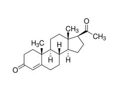
Biotium Progesterone(6-5E-10B), CF488A conjugate, 0.1mg/mL
This MAb is specific for progesterone. It exhibits minimal cross reactivity with related compounds in ELISA. It reacts with Progesterone-11a-HMS-BSA: 100%; 5-beta-Pregnane-3,20-dione: 48%; 5-alpha-Pregnane-3,20-dione: 26.4%; 17-alpha-Hydroxyprogesterone: 2.5% and 20-alpha-Hydroxyprogesterone: 0.04%. Progesterone is a steroid hormone synthesized from the cholesterol derivative, pregnenolone, in the cortex of the adrenal gland. Progesterone is secreted by the corpus luteum and acts to prepare the endometrium for the implantation of a fertilized egg. During pregnancy, it is secreted by the placenta to prevent spontaneous abortion and to stimulate the development of mammary tissue to produce milk. Thus, progesterone plays a central role in the reproductive events associated with the establishment and maintenance of pregnancy. Luteinized theca cells of normal ovary secrete progesterone. The determination of progesterone concentrations in the body fluids is of great value for endoc
Biotium Histiocytoma Marker(D11), CF488A conjugate, 0.1mg/mL
In Western blotting, this antibody detects an antigen of 125 kDa in human liver and 135 kDa in tumors of histiocytic origin. Comparative study of this MAb and a standard CD68 MAb showed that their antigens are different. Its antigen in all macrophage types studied is located on the plasma membrane and within cytoplasmic structures including lysosomes. This MAb shows a restricted reactivity to cells of the monocyte/macrophage system. It specifically reacts with blood monocytes and stains resident macrophages in a wide variety of human tissues. This MAb does not stain antigen-presenting cells, e.g., Langerhans cells. Reportedly, its reactivity is restricted to histiocytes and macrophages.Primary antibodies are available purified, or with a selection of fluorescent CF Dyes and other labels. CF Dyes offer exceptional brightness and photostability. Note: Conjugates of blue fluorescent dyes like CF405S and CF405M are not recommended for detecting low abundance targets, because
Biotium Histiocytoma Marker(D11), CF488A conjugate, 0.1mg/mL
In Western blotting, this antibody detects an antigen of 125 kDa in human liver and 135 kDa in tumors of histiocytic origin. Comparative study of this MAb and a standard CD68 MAb showed that their antigens are different. Its antigen in all macrophage types studied is located on the plasma membrane and within cytoplasmic structures including lysosomes. This MAb shows a restricted reactivity to cells of the monocyte/macrophage system. It specifically reacts with blood monocytes and stains resident macrophages in a wide variety of human tissues. This MAb does not stain antigen-presenting cells, e.g., Langerhans cells. Reportedly, its reactivity is restricted to histiocytes and macrophages.Primary antibodies are available purified, or with a selection of fluorescent CF Dyes and other labels. CF Dyes offer exceptional brightness and photostability. Note: Conjugates of blue fluorescent dyes like CF405S and CF405M are not recommended for detecting low abundance targets, because
Biotium HER-2 / CD340(HRB2/282), CF488A conjugate, 0.1mg/mL
Recognizes a protein of 185 kDa, which is identified as c-erbB-2/HER-2/neu. Its epitope is localized in the extracellular domain. C-erbB-2/HER-2 is a member of the EGFR family. This MAb is specific and shows minimal cross-reaction with other members of the EGFR-family. Receptors of this family are located on the plasma membrane and consist of an extracellular ligand-binding domain that is connected to a large intracellular domain by a single transmembrane sequence. c-erbB-2/HER-2 protein is over-expressed in a variety of carcinomas especially those of breast and ovary.Primary antibodies are available purified, or with a selection of fluorescent CF Dyes and other labels. CF Dyes offer exceptional brightness and photostability. Note: Conjugates of blue fluorescent dyes like CF405S and CF405M are not recommended for detecting low abundance targets, because blue dyes have lower fluorescence and can give higher non-specific background than other dye colors.
Biotium HLA-DRB(LN-3), CF488A conjugate, 0.1mg/mL
This MAb reacts with a 28 kDa chain of HLA-DRB1 antigen, a member of MHC class II molecules. It does not cross react with HLA-DP and HLA-DQ. The L243 antibody recognizes a different epitope than the LN3 monoclonal antibody, and these antibodies do not cross-block binding to each other's respective epitopes. HLA-DR is a heterodimeric cell surface glycoprotein comprised of a 36 kDa alpha (heavy) chain and a 28 kDa beta (light) chain. It is expressed on B-cells, activated T-cells, monocytes/macrophages, dendritic cells and other non-professional APCs. In conjunction with the CD3/TCR complex and CD4 molecules, HLA-DR is critical for efficient peptide presentation to CD4 T cells. It is an excellent histiocytic marker in paraffin sections producing intense staining. True histiocytic neoplasms are similarly positive. HLA-DR antigens also occur on a variety of epithelial cells and their corresponding neoplastic counterparts. Loss of HLA-DR expression is related to tumor microenviron
Biotium BCL-10(BL10/411), CF488A conjugate, 0.1mg/mL
B-cell lymphoma/leukemia 10 (BCL10) is a signaling protein that promotes apoptosis, pro-caspase-9 maturation, and activation of NF-kappa-B via NIK and IKK. BCL10 contains an N-terminal caspase recruitment domain (CARD), which is found in a number of apoptotic regulatory molecules. BCL10 is involved in the adaptive immune response, and may be an adapter protein between upstream TNFR1-TRADD-RIP complex and the downstream NIK-IKK-IKAP complex. A BCL10 translocation is recurrent in low-grade mucosa-associated lymphoid tissue (MALT lymphoma)Primary antibodies are available purified, or with a selection of fluorescent CF Dyes and other labels. CF Dyes offer exceptional brightness and photostability. Note: Conjugates of blue fluorescent dyes like CF405S and CF405M are not recommended for detecting low abundance targets, because blue dyes have lower fluorescence and can give higher non-specific background than other dye colors.